IV infiltration is the leakage of non-vesicant fluid from a peripheral intravenous catheter into the surrounding tissue, causing localized swelling, pain, and potential tissue damage. Severity ranges from minor discomfort to complications requiring surgical intervention.
This guide covers recognizing infiltration symptoms, responding when infiltration occurs, understanding risk factors and clinical assessment, treatment options, prevention strategies, and what to expect from a professional IV therapy provider.
Infiltration produces a distinct cluster of signs, including swelling, skin coolness, tightness, and pallor around the catheter site. Catching these changes early, through routine site inspection at least every eight hours, separates a quick catheter adjustment from a serious complication.
When infiltration is suspected, the immediate response follows a strict sequence: stop the infusion, leave the catheter in place for aspiration, avoid flushing the line, then elevate the limb. Patients discharged after an event should monitor for delayed reactions like blistering, worsening pain, or fever.
Certain populations face higher infiltration risk. Pediatric patients show PIVIE incidence rates near 17% in large observational data, while catheter indwelling time, insertion site, and device type all independently affect failure rates. Grading scales classify severity from no symptoms through blistering and absent pulses, and the assigned grade directly determines whether conservative care or emergency referral is needed.
Treatment ranges from elevation and agent-specific compresses for mild cases to pharmacological antidotes and surgical debridement for severe vesicant injuries. On the prevention side, smart infusion pumps, proper vein selection, and continuous site monitoring form the most reliable safeguards. Professional IV therapy providers apply these protocols during every session to minimize risk in real time.
What are the most common signs and symptoms of IV infiltration?
The most common signs and symptoms of IV infiltration are swelling, pain, skin coolness, and color changes at the IV insertion site. The sections below explain how to distinguish infiltration from other IV complications and which early warning signs to monitor during therapy.

How can you distinguish IV infiltration from other IV complications?
You can distinguish IV infiltration from other IV complications by comparing the specific cluster of symptoms present at the insertion site. Infiltration produces localized swelling, skin coolness, and a pale or blanched appearance because non-vesicant fluid accumulates in surrounding tissue. Phlebitis, by contrast, typically presents as warmth, redness, and a palpable cord along the vein. Hematoma causes visible bruising from blood pooling outside the vessel, while allergic reactions produce systemic signs such as hives or respiratory changes rather than localized coolness. According to St. Jude Children’s Research Hospital, IV infiltration signs include swelling, a tight or puffy feeling, pain, cool skin, and redness or pallor around the IV area. When multiple localized symptoms appear together without systemic involvement, infiltration is the most likely cause.
What are the early warning signs to watch for during IV therapy?
The early warning signs to watch for during IV therapy include subtle swelling or puffiness near the catheter insertion site, discomfort that was not present at the start of the infusion, and skin that feels cooler than surrounding areas. A slowed or stopped infusion flow, even without visible swelling, can also signal that fluid is no longer entering the vein properly. Because these changes develop gradually, routine site checks are essential. The Australian Commission on Safety and Quality in Health Care recommends inspecting the peripheral IV catheter and insertion site at least once every eight hours and whenever the device is accessed. Catching even mild tightness or a slight temperature drop early allows prompt intervention before tissue damage progresses.
What immediate steps should you take if IV infiltration is suspected?
The immediate steps you should take if IV infiltration is suspected include stopping the infusion, preserving the IV access device for aspiration, and elevating the affected limb. The sub-sections below cover clinical response protocols and at-home aftercare strategies.

What actions should healthcare providers perform right after detecting infiltration?
Healthcare providers should perform a structured sequence of actions right after detecting infiltration. According to the Cleveland Clinic Journal of Medicine, the recommended steps are:
- Stop administration of fluid immediately.
- Disconnect the IV tubing, but leave the catheter in place to allow aspiration of leaked fluid.
- Do not flush the line.
- Aspirate as much infiltrated fluid as possible through the catheter.
- Remove the catheter if no antidote will be administered through it.
- If an antidote is indicated, inject it through the catheter before removal to ensure delivery to the extravasation site.
- Elevate the affected limb and apply warm or cold compresses based on the infused agent.
Because severity depends on the volume infused and the drug’s pH, osmolarity, and pharmacological action, prompt aspiration minimizes the risk of tissue necrosis or compartment syndrome. Delaying even one of these steps can mean the difference between a minor complication and a surgical referral.
What at-home care strategies can help after an infiltration event?
At-home care strategies that can help after an infiltration event focus on monitoring, wound protection, and knowing when to seek urgent follow-up. According to the Oncology Nursing Society’s extravasation guidelines, patients should monitor the area for redness, pain, blistering, peeling, or streaks, and report delayed reactions such as fever higher than 100.4°F. Key at-home strategies include:
- Keep the affected area clean, dry, and free from tight clothing or pressure.
- Elevate the limb to reduce swelling.
- Apply warm or cold compresses as directed by the treating clinician (compress choice is agent-specific).
- Watch for worsening pain, new blistering, diminished range of motion, or changes in sensation.
Severe extravasation can develop hours after the initial event, so vigilance at home is not optional. Recognizing these risk factors early connects directly to understanding which patients face higher infiltration risk overall.
What are the risk factors and causes associated with IV infiltration?
The risk factors and causes associated with IV infiltration span patient-specific vulnerabilities and procedural variables. The sections below cover susceptible patient populations and how equipment and technique choices influence infiltration risk.
Which patient populations are more susceptible to IV infiltration?
The patient populations more susceptible to IV infiltration include pediatric patients, older adults, and individuals with fragile or compromised vasculature. Children present smaller, more mobile veins that are harder to stabilize during catheter placement. Older adults often have thinner skin and decreased vein elasticity, which increases the likelihood of catheter dislodgement.
According to a retrospective observational study published in the Journal of International Medical Research examining 11,006 pediatric surgery inpatients and 19,771 peripheral intravenous catheters (PIVCs), the incidence of peripheral intravenous therapy infiltration and extravasation (PIVIE) reached 16.93%. Significant predictors included patient age, sex, disease classification, puncture site, and indwelling time. Clinicians should recognize that nearly one in six pediatric PIVCs in that study experienced infiltration or extravasation, underscoring the need for heightened vigilance in younger patients.

How do equipment type and technique influence infiltration risk?
Equipment type and technique influence infiltration risk by affecting catheter stability, insertion accuracy, and fluid delivery pressure. Catheter gauge, insertion site selection, securement method, and the type of vascular access device all play measurable roles.
Key equipment and technique factors include:
- Catheter gauge and length: Oversized catheters relative to vein diameter increase mechanical irritation and raise failure rates.
- Insertion site: Joints and areas of high mobility, such as the antecubital fossa or wrist, promote catheter migration.
- Securement quality: Inadequate dressing or taping allows the catheter tip to shift within or outside the vein.
- Indwelling time: Longer dwell periods correlate with higher PIVIE incidence, as tissue and vessel integrity degrade around the catheter.
- Vascular access device type: A randomized clinical trial published in JAMA Network Open found that midline catheters had a 13.2% complication rate compared with 7.2% for peripherally inserted central catheters (PICCs), with an incidence rate ratio of 2.37 for complications.
Proper catheter selection, secure fixation, and routine site assessment remain the most controllable variables for reducing infiltration risk. Understanding these factors helps clinicians and patients recognize when procedural choices elevate infiltration likelihood, which informs the diagnostic methods covered next.
How is IV infiltration diagnosed and assessed?
IV infiltration is diagnosed and assessed through clinical examination of the IV site combined with standardized grading scales that classify severity. The following subsections cover the diagnostic methods clinicians use and how severity grades guide treatment decisions.
What diagnostic methods and grading scales are used for IV infiltration?
The diagnostic methods used for IV infiltration include visual inspection, palpation of the catheter insertion site, and standardized severity grading scales. According to the CDC’s Guidelines for the Prevention of Intravascular Catheter-Related Infections, clinicians should “evaluate the catheter insertion site daily, by palpation through the dressing to discern tenderness and by inspection if a transparent dressing is in use.”
The most widely referenced grading tool is the Infiltration Scale, which classifies events from Grade 0 (no symptoms) through Grade 4 (severe). Key indicators assessed at each grade include:
- Swelling diameter and location relative to the insertion site
- Skin temperature changes, such as coolness to the touch
- Blanching or skin discoloration around the IV area
- Pain intensity at or near the catheter site
- Pulse quality and capillary refill time distal to the site
At the highest severity level, clinicians look for skin blistering, necrosis, absent pulse, and capillary refill exceeding four seconds. Consistent use of a grading scale standardizes documentation and ensures appropriate escalation.
How does the severity of infiltration affect treatment decisions?
The severity of infiltration directly determines the urgency and type of treatment a patient receives. Low-grade infiltration involving minor swelling and no tissue damage typically requires stopping the infusion, removing the catheter, and applying elevation with a compress. These cases often resolve without further intervention.
Higher-grade infiltration demands a more aggressive response. According to St. Jude Children’s Research Hospital, signs like blistering, necrosis, and significant pain warrant immediate medical evaluation and possible surgical consultation. When blanching, absent pulses, or capillary refill beyond four seconds are present, the risk of compartment syndrome or permanent tissue damage increases substantially.
For clinicians, grading severity at the point of detection is not optional; it is the decision framework that separates a simple site change from an emergency referral.
Accurate assessment at the time of detection shapes every downstream treatment choice, from conservative management to specialist intervention.
What are the most effective treatments for IV infiltration?
The most effective treatments for IV infiltration include both pharmacological antidotes and non-pharmacological interventions such as elevation and compresses. The approach depends on the substance involved and injury severity.
What pharmacological and non-pharmacological interventions are recommended?
The pharmacological and non-pharmacological interventions recommended for IV infiltration depend on the extravasated agent and severity of tissue injury.
Non-pharmacological measures form the foundation of treatment:
- Elevate the affected limb to reduce swelling and promote fluid reabsorption.
- Apply warm or cold compresses based on the specific infusate involved.
- Keep the area clean and dry while avoiding tight clothing over the site.
For vesicant extravasation, pharmacological antidotes target specific drug classes. According to the University of Illinois Chicago Drug Information Group, hyaluronidase is used for extravasation of vinca alkaloids, taxanes, etoposide, hyperosmolar agents, and agents containing propylene glycol, with 15 to 25 units administered intradermally over 5 injections, ideally within 1 hour of the event.
When conservative therapy fails and tissue necrosis or pain persists beyond 10 days, surgical debridement followed by skin grafting may be required. Early, agent-specific intervention is what separates a minor complication from a serious wound.
How long does recovery from IV infiltration typically take?
Recovery from IV infiltration typically takes a few days for mild cases, though severe extravasation injuries may require weeks of monitoring or longer if surgical intervention becomes necessary.
Mild infiltration involving non-vesicant fluids generally resolves within 24 to 72 hours with elevation and compress application. Vesicant-related injuries follow a less predictable timeline because, as noted by eviQ, early vesicant extravasation signs may be subtle and not always evident until days or weeks later. For this reason, daily or second-daily clinical review is recommended during the first week, followed by weekly review until complete resolution.
Patients discharged after an infiltration event should watch for delayed warning signs, such as worsening pain, new blistering, or decreased range of motion, and seek immediate care if these develop.
Understanding treatment options sets the stage for exploring how infiltration can be prevented altogether.
How can IV infiltration be prevented during infusion therapy?
IV infiltration can be prevented during infusion therapy through a combination of clinical best practices and monitoring technologies. The following subsections cover practical nursing techniques and device-based safeguards that reduce peripheral intravenous therapy infiltration and extravasation (PIVIE) risk.
What best practices can reduce the likelihood of IV infiltration?
Best practices that reduce the likelihood of IV infiltration include proper site selection, secure catheter stabilization, and routine monitoring throughout the infusion. The following strategies form the foundation of PIVIE prevention:
- Select an appropriate vein, favoring the forearm over the hand or wrist, where joint movement increases dislodgement risk.
- Use the smallest gauge catheter suitable for the prescribed therapy to minimize vessel trauma.
- Secure the catheter with a transparent dressing so the insertion site remains visible for continuous inspection.
- Inspect the insertion site at regular intervals during infusion, checking for swelling, coolness, or pain.
- Educate patients to report tightness, burning, or discomfort at the IV site immediately.
- Limit catheter indwelling time, since longer dwell periods independently increase infiltration risk.
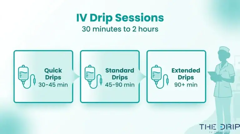
For wellness infusions specifically, where sessions typically last around one hour, consistent visual and tactile site checks during that window are highly practical and often sufficient to catch early changes before tissue damage occurs.

What technologies can help prevent infiltration during IV infusions?
Technologies that help prevent infiltration during IV infusions include smart infusion pumps, pressure-sensing alarms, and near-infrared vein visualization devices. According to a systematic review published by the Agency for Healthcare Research and Quality (AHRQ) Patient Safety Network, smart infusion pumps with pre-programmed dosing libraries can effectively prevent medication administration errors and clinical adverse drug events.
Key technology-based safeguards include:
- Smart infusion pumps that detect pressure changes in the IV line and trigger alarms when resistance suggests fluid is no longer flowing into the vein.
- Near-infrared vein finders that improve first-stick accuracy by illuminating subcutaneous vasculature, reducing the need for multiple insertion attempts.
- Transparent, securement-integrated dressings that allow continuous visual monitoring without disturbing catheter position.
No single device eliminates infiltration entirely. Pairing these technologies with trained clinical oversight remains the most reliable prevention approach.
Understanding prevention strategies helps set realistic safety expectations when choosing any IV therapy provider.
How should you approach IV infiltration concerns with The Drip IV Infusion?
You should approach IV infiltration concerns with The Drip IV Infusion by understanding how trained infusion specialists minimize risk and by knowing what signs warrant immediate attention. The sections below cover prevention support and a summary of the key infiltration insights discussed throughout this article.
Can The Drip IV Infusion help prevent or manage IV infiltration?
Yes, The Drip IV Infusion can help prevent IV infiltration by having experienced infusion specialists administer every session. Founded by professionals with decades of nursing and emergency medicine backgrounds, The Drip IV Infusion uses trained nurses who follow established catheter management practices, including regular site monitoring during each one-hour session.
According to an NHS Greater Glasgow and Clyde neonatal guideline on extravasation injuries, severe infiltration requires immediate infusion cessation, aspiration of fluid from the catheter, and prompt medical review before device removal. While wellness IV sessions involve non-vesicant fluids with lower complication risk, knowing these escalation protocols matters. The Drip IV Infusion offers both mobile and in-office treatments across Arizona, so clients can raise concerns directly with their infusion specialist in real time.
For any unusual swelling, coolness, or pain at the IV site, always speak up immediately.
What are the key takeaways about IV infiltration signs, treatment, and prevention we covered?
The key takeaways about IV infiltration signs, treatment, and prevention are:
- Recognize early signs promptly. Swelling, skin coolness, pain, and tightness around the IV site all signal possible infiltration and require immediate attention.
- Stop the infusion first. Halting fluid delivery and notifying the clinician is the single most important initial step before any further intervention.
- Treatment depends on severity and the infused substance. As the University of Illinois Chicago Drug Information Group notes, specific antidotes exist for certain drug extravasations, and timing is critical; ideally, treatment begins within one hour of the event.
- Prevention starts with skilled catheter placement. Proper vein selection, regular site checks, and experienced clinicians significantly reduce infiltration risk.
- Patient awareness is protective. Reporting discomfort early leads to faster detection and better outcomes.
Understanding these fundamentals helps you approach any IV therapy session, whether at home or in a clinical setting, with greater confidence and safety awareness.